

SAFA
ASSESSMENT
ASSESSMENT
Skolkovo & Clinic
SAFA Facial Assessment for Doctors
Dr Hotaki’s framework for assessing anatomy, proportions and tissue displacement. SAFA is studied within the training programme, alongside UPS planning and clinical practice.
A maximum of four doctors per group
Training for Aesthetic Physicians
Aesthetic medicine
does not begin with a procedure
does not begin with a procedure
SAFA provides a structured approach to facial assessment before deciding whether and how to intervene.
Because technical skill alone is not enough. A doctor must understand:
What has changed in the patient’s face
Where tissue displacement has occurred
01
02
How pronounced the changes are
03
The direction in which the tissues have moved
04
Which anatomical structures require support
The safe anatomical plane for implant placement
05
06
Which vector will achieve natural tissue repositioning
07
Where the fixation point should be located
08
When the procedure is indicated
09
When a different treatment should be selected
10
When an aesthetic intervention should not be performed
11
During the programme, you do not simply learn a standardised pattern. You develop a structured process of clinical reasoning:
Why should two patients with similar concerns not receive the same treatment plan?
Why does one vector genuinely reposition tissue while another creates only superficial tension?
Why does a technically correct implant placement not always produce an aesthetically correct result?
- ASSESS

2. CLASSIFY

3. PLAN

4. POSITION

5. PERFORM

6. REASSESS

MODULE
CONTENT
CONTENT
Six aspects of SAFA: structured assessment, tissue displacement, proportions, anatomy, clinical planning and its relationship to UPS.
01
02
03
04
05
STRUCTURED ASSESSMENT
Record anatomy, proportions and tissue distribution before considering a treatment plan.
TISSUE DISPLACEMENT
Assess anatomy, tissue distribution and facial proportions before planning treatment.
PROPORTIONS AND ASYMMETRY
Examine facial relationships and asymmetry as part of individual assessment.
ANATOMICAL RELATIONSHIPS
Relate visible changes to underlying tissues and relevant anatomical structures.
CLINICAL PLANNING
Use assessment findings to discuss indications, limitations and individual planning.
There are contraindications. Consultation with a specialist is required. Results vary individually and are not guaranteed.
SAFA is studied within the author-led training programme. It is not a separate five-day course.
06
SAFA AND UPS
SAFA concerns assessment. UPS supports subsequent positioning and vector planning.
Structured assessment


Anatomy and proportions

Individual planning

Clinical reasoning

The five-day programme below provides the training context in which SAFA is studied. SAFA is not a separate five-day module.



Five days,
that transform clinical thinking
that transform clinical thinking
DAY 1
ASSESSMENT AND PLANNING
The first day establishes the foundation for all subsequent training
• the history and development of the method;
• the structure and properties of the implant;
• the design of the proprietary needle;
• introduction to SAPA and SAFA;
• standardised patient assessment;
• diagnosis of ptosis;
• evaluation of tissue displacement;
• classification of asymmetry;
• the UPS system;
• construction of individualised vectors;
• identification of fixation points;
• indications and limitations;
• patient selection;
• principles of local anaesthesia;
• principles of preparing a tumescent solution;
• clinical photography protocols;
• informed consent;
• standardised clinical documentation.
The programme includes:
DAY 2
TOPOGRAPHICAL ANATOMY AND CADAVERIC DISSECTION
Training is delivered with the involvement of experts and professors of anatomy
The objective of Day 2 is to teach doctors to see beyond the surface and understand the structures through which, or close to which, the instrument passes.
• layered anatomy of the face or selected treatment area;
• fasciae;
• retaining ligaments;
• fat compartments;
• anatomical spaces;
• arteries;
• veins;
• nerves;
• high-risk anatomical zones;
• the temporal region;
• fixation points;
• anatomical justification of the implant trajectory;
• cadaveric dissection.
• fasciae;
• retaining ligaments;
• fat compartments;
• anatomical spaces;
• arteries;
• veins;
• nerves;
• high-risk anatomical zones;
• the temporal region;
• fixation points;
• anatomical justification of the implant trajectory;
• cadaveric dissection.
The programme includes:
DAY 3
AUTHOR-LED DEMONSTRATION AND ULTRASOUND GUIDANCE
Dr Hotaki demonstrates the proprietary technique on cadaveric material
Ultrasound enables participants to connect theoretical anatomy and cadaveric dissection with the actual position of the implant.
• the actual anatomical plane of the implant;
• its relationship to surrounding structures;
• the correct placement trajectory;
• changes in depth across different anatomical regions;
• potential technical deviations;
• criteria for safe performance of the procedure.
• its relationship to surrounding structures;
• the correct placement trajectory;
• changes in depth across different anatomical regions;
• potential technical deviations;
• criteria for safe performance of the procedure.
Using ultrasound visualisation, participants observe:

DAY 4
SUPERVISED PRACTICAL TRAINING
The implant plane and trajectory are subsequently verified using ultrasound.
Errors are not simply corrected. Their causes and potential clinical consequences are analysed in detail.
Each participant performs the principal stages of the method on cadaveric material under direct supervision
• handling the proprietary needle;
• selecting the entry point;
• controlling the direction of movement;
• following the planned trajectory;
• reaching the required anatomical plane;
• creating the tissue-support system;
• working with fixation points;
• following the safety algorithm.
• selecting the entry point;
• controlling the direction of movement;
• following the planned trajectory;
• reaching the required anatomical plane;
• creating the tissue-support system;
• working with fixation points;
• following the safety algorithm.
Doctors practise:
DAY 5
CLINICAL DEMONSTRATION ON LIVE MODELS
The final day takes place in a licensed clinic
• medical history-taking;
• patient assessment using SAFA;
• identification of indications and limitations;
• standardised clinical photography;
• construction of the UPS markings;
• vector selection;
• identification of fixation points;
• patient preparation;
• performance of the procedure by Dr Hotaki;
• explanation of each clinical decision;
• post-procedure management;
• patient aftercare instructions;
• outcome assessment and documentation.
It brings together every component of the programme:

The five-day programme creates one integrated pathway
from the initial patient assessment to safe performance and subsequent monitoring

Frequently asked questions:
Standardised Aesthetic Facial Assessment is Dr Hotaki’s author-developed framework for structured facial assessment before clinical planning. Read the history of SAFA and the clinical need behind its development. Why I believe my training is a revolution in aesthetic medicine.
Anatomy, facial proportions, asymmetry, tissue distribution and displacement, and their relevance to treatment planning.
SAFA concerns assessment. The Universal Positioning System supports subsequent positioning and vector planning. They address different stages of the clinical process.
No. SAFA is studied within the author-led training programme, including face lift and neck lifting modules. See also brow lifting, upper lip lifting and non-surgical alarplasty training.
This page describes Dr Hotaki’s author-developed assessment framework. It does not present SAFA as a universally recognised or independently validated scientific standard.


Your next procedure should not begin with the choice of an implant.
Places are limited: each group includes a maximum of four doctors.
Request the programme that includes SAFA.
the complete programme for your chosen direction;
upcoming training dates;
information about joining the graduate community.
a list of the documents awarded;
fees and booking terms;
information about the training venue;
participant requirements;
professional selection criteria;
A maximum of four doctors per group.
Four doctors. Five days. Maximum focus on the development of every participant.
This is not high-volume training or a demonstration masterclass for a large audience.
No participant remains merely an observer
Each training group is limited to a maximum of four doctors. This small-group format allows Dr Hotaki to personally oversee the development of every participant:
Each training group is limited to a maximum of four doctors. This small-group format allows Dr Hotaki to personally oversee the development of every participant:

answer individual questions.

examine complex clinical situations in detail;

confirm understanding of anatomical planes;

identify and analyse technical errors;
evaluate handling of the proprietary needle;
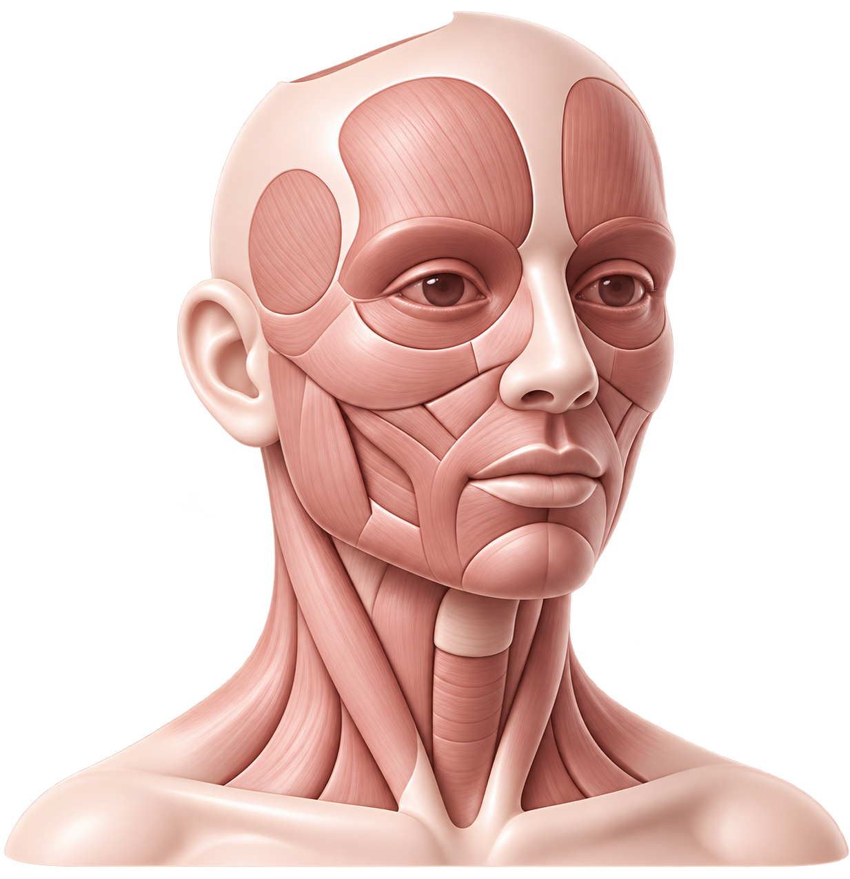
supervise work on cadaveric material;
review vector selection;

refine patient analysis;
assess clinical reasoning;
Strict professional selection.
We assess:
medical education;
professional motivation.
readiness to maintain standardised clinical documentation;
readiness to follow safety requirements;
experience with injectable or thread-based procedures;
knowledge of anatomy;
experience in aesthetic medicine;
clinical experience;
current professional documentation;
professional qualifications;
Places are limited to preserve the quality and safety of the training — not to create artificial scarcity.
A preliminary interview with Dr Hotaki may also be required.
All candidates therefore undergo a preliminary selection process before admission.
Admission to the programme is not automatic. These proprietary methods require a detailed understanding of anatomy, a responsible approach to patient safety and a commitment to following clinical protocols.
The purpose of selection is to form a small group of doctors who are prepared not merely to observe the technique, but to learn the complete proprietary clinical system.

Documentation awarded on completion.
Following successful completion of training, the doctor may offer the relevant procedure to patients, provided that they hold the necessary professional qualifications and comply with the regulatory requirements of the country in which they practise.
These documents confirm that the doctor has completed the theoretical, anatomical and practical stages of the programme and has learned the proprietary protocol for the selected direction.
Following successful completion of each individual direction, the doctor receives:
A named certificate confirming completion of training in Dr Hotaki’s proprietary method;

A diploma confirming completion of the relevant educational programme;

A certificate issued in accordance with the applicable state educational standard;
Registration of the awarded document in the relevant state register.
Completion is determined not simply by attendance, but by demonstrated achievement of the programme’s requirements.
Clinic address


115114 Moscow, Derbenevskaya Embankment 11,
BC POLLARS
BC POLLARS
Contacts

Training for cosmetic doctors in a patent-pending permanent lifting technique
Specialized modules, anatomy with professors, ultrasound guidance, and only four places per cohort
© 2026. All rights reserved
For medical professionals only. Contraindications apply.