Guided
Permanent Lifting


At the core of the patent-pending technique is a flat,
non-absorbable implant and an original method of fixation
A Continuing Education Diploma
and 36 Credit Points
The training program is approved under state education standards for the accreditation of aesthetic physicians. Documents are issued by the Moscow Medical Institute.
Each treatment area is five days of training. Physicians use these points toward their next accreditation cycle.
Certificate and Continuing Education Diploma
State-format documents are issued by the Moscow Medical Institute. Physicians separately receive a personalized certificate bearing Dr. Hotaki's own signature.
Entry in the State Register
Details of issued documents are entered into the state register, so the training is officially verifiable — not confirmed only within the program itself.
Counts Toward Accreditation
Physicians present the points earned for completed treatment areas during their next accreditation, so training in the technique isn't separate from that process.
and dermatologist in Russia

in Aesthetic Medicine


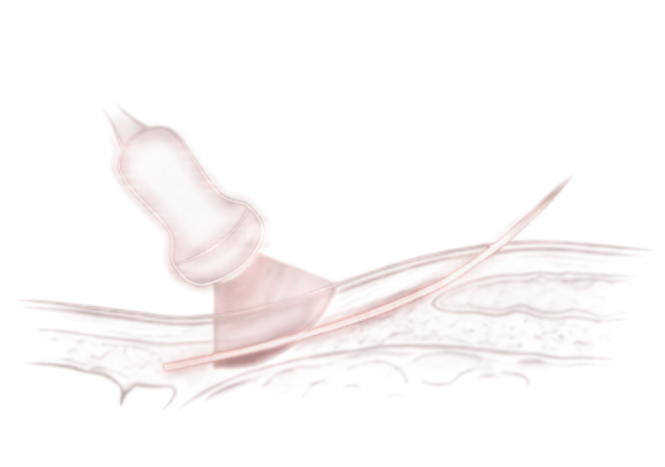
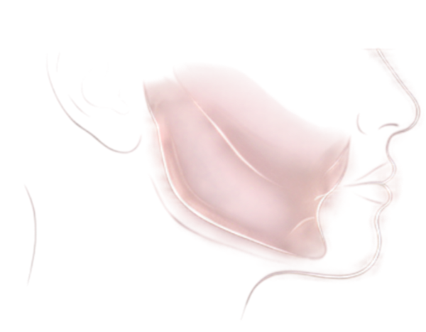
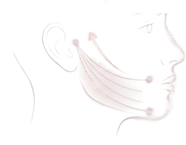
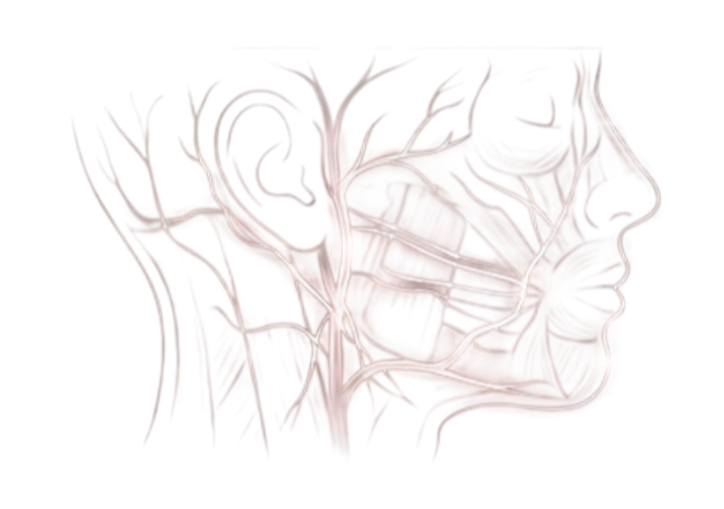
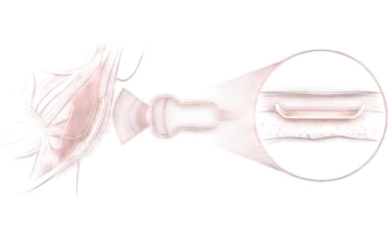

isn't taught by video
technique control



Patient Selection

for the procedure

с привычными решениями
подтяжка
в местах разрезов
нити
за 6-18 месяцев
Доктора Хоттаки
доступ через прокол
с распределением нагрузки

и курс по чужой методике

of the original technique


per treatment area









- Technique


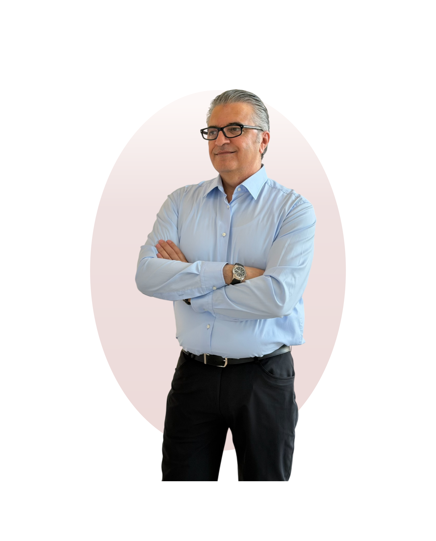
Dr. Nour Hotaki


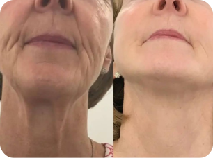
лифтинг лица и шеи
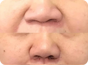
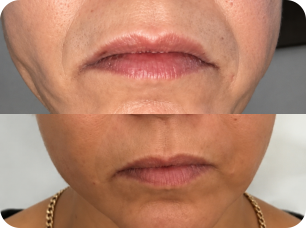
нитевого лифтинга
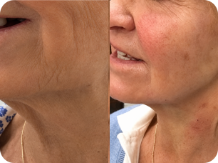
лифтинг лица и шеи
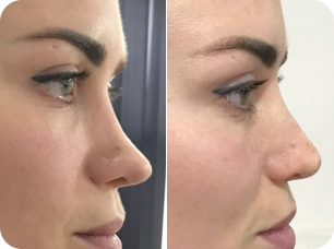
ринопластики

that is 24 procedures per year




by Dr. Hotaki